


Synthetic bone substitutes and collagen membranes in dentistry
Reconstructive bone surgery aims to regenerate the loss or resorption of bone through materials and techniques that allow to mimic and activate specific and fundamental reparative mechanisms such as osteogenesis, osteoinduction, and osteoconduction.
- Osteogenesis: formation of new bone tissue by the direct action of progenitor cells and osteoblasts
- Osteoinduction: regulatory activity of growth factors capable of triggering the differentiation of mesenchymal cells in the osteoblastic lineage and the process of bone regeneration
- Osteoconduction: support provided by a scaffold that allows the deposition of a new osteoid substance
Any regenerative technique must comply with these three elements, together with the vascularization of the treated site and the stability of the graft. When the bone defects are small and the patient is in good health, the bone has an excellent healing capacity, with intrinsic “genesis” and “induction”; it is sufficient that the surgeon fills the void of the loss of substance with grafts or bone substitutes to promote only osteoconduction. This provides the three-dimensional structure to sustain the regeneration process.
Autologous bone is taken from the patient himself and offers many advantages because, in addition to the osteoconductive bone matrix, it can also provide osteogenic and osteoinductive elements thanks to the presence of cells and growth factors. However, the availability of autologous bone in the endo-oral site is limited, while harvesting from other parts of the body (iliac bone) is invasive, risky, and not always possible. In addition to autologous tissue grafting, there are three main categories of bone substitutes: homologous, heterologous, and synthetic.
Homologous bone is from a cadaveric donor. It can be received from tissue banks, but even in this case, various limitations do not always allow its use.
Heterologous bone substitutes are biological grafts derived from animals (usually bovine or equine) that are decellularized and cleared of all antigenic elements. They offer different solutions, in granules or blocks of cortical, or spongy bone, with different resorption and revascularization times. Some heterologous bone substitutes detain a preserved protein/collagen component that can promote rapid osseointegration, as well as indirect osteoinduction.
Synthetic bone substitutes, available in a wide range of different materials, e.g., hydroxyapatites, calcium phosphates and sulfates, have very heterogeneous characteristics usually provide the osteoconductive scaffold for bone neoformation.
SpherHA synthetic bone substitutes
Tiss’You has developed SpherHA, a line of synthetic bone substitutes, composed of bio-mimetic nano-structured hydroxyapatite. Hydroxyapatite is a mineral found in the inorganic component of human bone. Both the composition and crystal structure of SpherHA are extremely similar to the mineral matrix of our bones, with a calcium to phosphate ratio of 1.67: the same as natural bone apatite.
 The highly porous and interconnected structure and its composition ensure an excellent osteoconduction and circulation of nutrients, and also a real active stimulus to the formation of new bone tissue, favoring the processes of cell colonization and early vascularization. The nano-structure of SpherHA crystals provides a very high surface area to volume ratio that ensures complete degradation by osteoclastic activity with consequent remodeling into new viable bone tissue. These elements, along with the bio-mimetic properties, make SpherHA an ideal bone substitute for osseointegration and regeneration of bone defects.
SpherHA is available in several formats: porous chips, dense granules, injectable paste, and moudable crunch. In dental surgery, the indication is for filling small and medium periodontal and peri-implant bone defects, for filling post-extraction sockets, and for sinus lift.
Download SpherHA catalog (PDF)
Collagen and GBR (Guided Bone Regeneration)
In reconstructive bone surgery, especially in dentistry, it is important to guide bone growth limited to the damaged area, using barrier membranes to prevent the entry of not-osteogenic elements in the treated site. This procedure, called GBR (Guided Bone Regeneration), avoids the infiltration of fibroblasts from adjacent soft tissues, thus preventing the formation of fibrous tissue at the graft site.
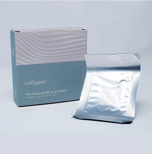 For this purpose, Tiss’You has developed Collygen, an absorbable membrane of equine-derived atelocollagen, easily applicable without the need for fixation, for the protection of bone grafts in regenerative surgery techniques. Collygen, thanks to its collagen texture, actively participates in the healing process of the treated site, in addition to its passive protection activity. Recently, it has been demonstrated that the collagen membranes, which in the past were considered only passive barriers, act instead as a real bioactive compartment, interacting with the cellular components that home to the graft site. The processes thus activated contribute to the formation of bone tissue and its improved remodeling.